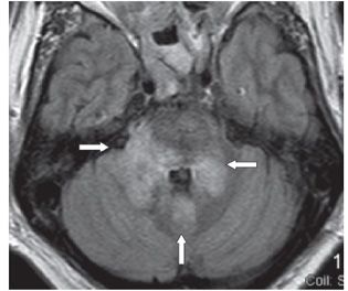
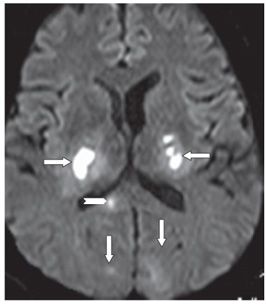
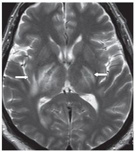
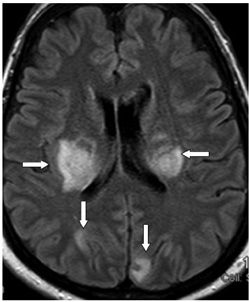
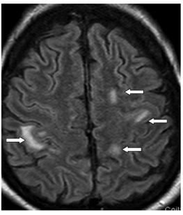
FINDINGS Figures 46-1 and 46-2. Axial DWI and FLAIR MRI through the brachium pontes. There are central areas of diffusion restriction within T2 hyperintensity in bilateral brachium pontes extending into adjacent pons and cerebellum bilaterally (transverse arrows). The nodulus also shows tiny restricted diffusion within a larger T2 hyperintensity (vertical arrows). Figures 46-3 and 46-4. Axial DWI and T2WI through the thalami. There are bilateral thalamocapsular and posterior lentiform nuclei areas of diffusion restriction within larger areas of T2 hyperintensity possibly surrounding edema (arrows). There are smaller lesions in the bilateral subcortical occipital lobes (vertical arrows) and the splenium (chevron) in Figure 46-3. Images of the midbrain (not shown) revealed similar changes. Figure 46-5. Axial FLAIR images through the corona radiata. There is almost bilateral symmetrical coronal radiata periventricular (transverse arrows) and occipital subcortical (vertical arrows) hyperintensity. Figure 46-6
Stay updated, free articles. Join our Telegram channel

Full access? Get Clinical Tree


