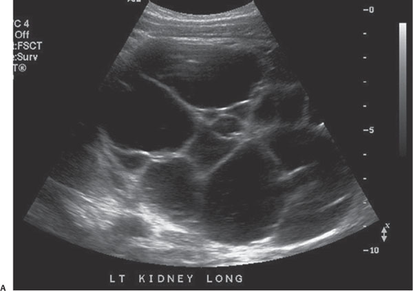
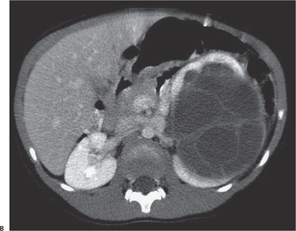
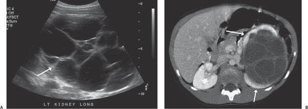
Case 46 A 6-month-old boy with a palpable abdominal mass. (A) Longitudinal ultrasound image: There is a multicystic mass in the left kidney (arrow). The cysts do not appear to communicate. (B) Contrast-enhanced computed tomography (CT) image: There is a multiloculate, fluid-density mass herniating into the left renal collecting system. Normally enhancing kidney surrounds it (arrows).
Clinical Presentation
Further Work-up
Imaging Findings
Differential Diagnosis
Stay updated, free articles. Join our Telegram channel

Full access? Get Clinical Tree


