 Clinical Presentation
Adult patient with severe chest pain and worsening subcutaneous emphysema after placement of chest tube for left-sided pneumothorax.
 Imaging Findings
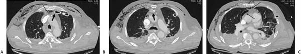
(A–C) Computed tomography (CT) of the chest: Axial images at three different levels demonstrate a left-sided thoracostomy tube with its tip within the fat of the anterior mediastinum (thick black arrows), as well as a significant degree of pneumomediastinum and subcutaneous emphysema (thin black arrows). Bilateral parenchymal opacities are also noted.
 Differential Diagnosis
• Malposition of a thoracostomy tube:
Stay updated, free articles. Join our Telegram channel

Full access? Get Clinical Tree


