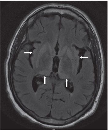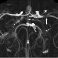
FINDINGS Figures 47-1 and 47-2. Axial DWI, and corresponding FLAIR at the level of basal ganglia. Diffusion restriction and increased FLAIR signal are noted in the bilateral corpus striatum (transverse arrows) and thalamus (vertical arrows). Two characteristic signs—pulvinar sign (symmetric involvement of pulvinar, the posterior nuclei of thalamus) and “hockey stick” sign (symmetric involvement of pulvinar and dorsomedial thalamus)—are present (vertical arrows). There is also involvement of the insula cortex.
DIFFERENTIAL DIAGNOSIS Acute hypoxic ischemic encephalopathy (HIE), encephalitis, other causes of dementia (Alzheimer disease, frontotemporal dementia, multi-infarct dementia, corticobasal degeneration), Leigh syndrome, Wilson disease, Creutzfeldt-Jakob disease.
DIAGNOSIS Creutzfeldt-Jakob disease (CJD), variant form.
DISCUSSION
Stay updated, free articles. Join our Telegram channel

Full access? Get Clinical Tree


