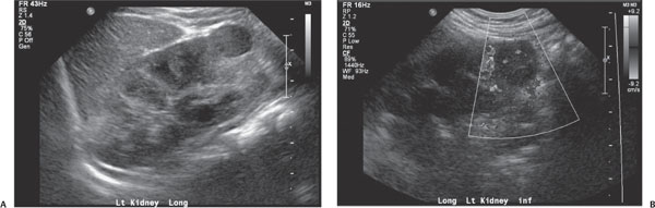
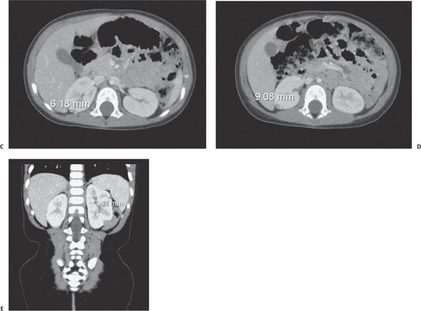
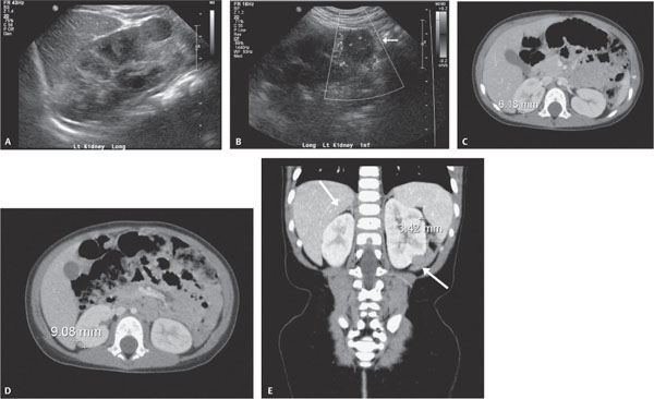
Case 47 A 3-year-old boy undergoing ultrasound for abdominal pain. (A,B) US: There is a hypoechoic round mass at the lower pole of the left kidney with internal blood flow (arrow). (C–E) Computed tomography (CT): There are small, homogeneous, low-density nodules in the periphery of the kidneys (arrows). They do not enhance as strongly as the normal renal cortex. • Nephroblastomatosis: Multiple small, solid, homogeneous renal masses are consistent with nephroblastomatosis. • Wilms tumor:
Clinical Presentation
Further Work-up
Imaging Findings
Differential Diagnosis
![]()
Stay updated, free articles. Join our Telegram channel

Full access? Get Clinical Tree


