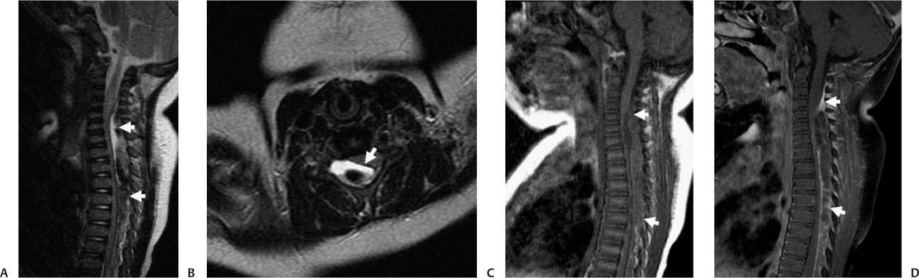
Case 48 A 1-year-old with the sudden onset of bilateral lower extremity weakness. (A) Sagittal T2-weighted image (WI) shows a biconcave collection in the lower cervical and upper thoracic dorsal epidural space (arrows) with heterogeneous signal and areas of low signal. (B) In this axial image, the dura is visualized as a linear structure with low signal (arrow) between the anteriorly located cord and the hematoma. Note the compressive effect on the cord, which does not show edema. (C,D) Sagittal T1WIs, pre- and postcontrast administration, show scattered linear areas of enhancement (arrows) within and at the periphery of the collection. • Epidural hematoma (EDH): EDH is an extra-axial collection of blood with heterogeneous signal intensity and a biconvex shape. Displacement of the dura toward the cord confirms the epidural location of the lesion. • Epidural abscess:

Clinical Presentation
Imaging Findings
Differential Diagnosis
![]()
Stay updated, free articles. Join our Telegram channel

Full access? Get Clinical Tree


