 Clinical Presentation
Chest radiograph from an adult patient after a cardiac intervention.
 Imaging Findings
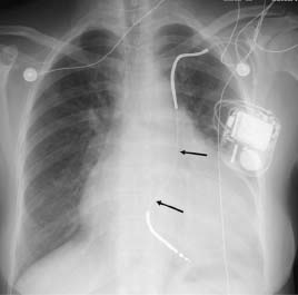
Single anteroposterior (AP) radiograph of the chest shows the lead of the pacing device coursing from a left subclavian approach to the left of midline and terminating in the usual position for the right ventricle (arrows). A significantly enlarged cardiac silhouette with a left ventricular configuration is also appreciated.
 Differential Diagnosis
• Persistent left superior vena cava (PLSVC): PLSVC is typically found incidentally on a chest radiograph obtained after a placement of a left-sided central venous line or during contrast-enhanced computed tomography. Occasionally, a conventional chest radiograph may show mild mediastinal widening with a soft-tissue density to the left of the aortic knob. On cross-sectional images, the PLSVC is seen as a vascular structure connecting the confluence of the left subclavian vein and internal jugular vein with the coronary sinus.
• Partial anomalous pulmonary venous return (PAPVR):
Stay updated, free articles. Join our Telegram channel

Full access? Get Clinical Tree


