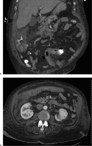
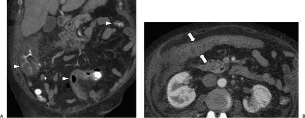
Case 48 A 55-year-old man being treated for a kidney infection presents with diarrhea. (A) Contrast-enhanced computed tomography (CT) shows marked, diffuse circumferential thickening of the colon wall (arrowheads), trapped barium in mucosal folds, and mesenteric fat stranding. (B) Axial image shows circumferential thickening of the transverse colon (arrows) with obliteration of the lumen. • Infection: This is the most likely diagnosis, given that this patient has pseudomembranous colitis. Marked, diffuse, symmetric circumferential colon wall thickening with barium trapped in thick haustral folds is most typical of infections such as those caused by Clostridium difficile or cytomegalovirus (CMV; seen in acquired immunodeficiency syndrome). Also consider Salmonella, Shigella, or Campylobacter infection. • Inflammation:
 Clinical Presentation
 Imaging Findings
 Differential Diagnosis
![]()
Stay updated, free articles. Join our Telegram channel

Full access? Get Clinical Tree


