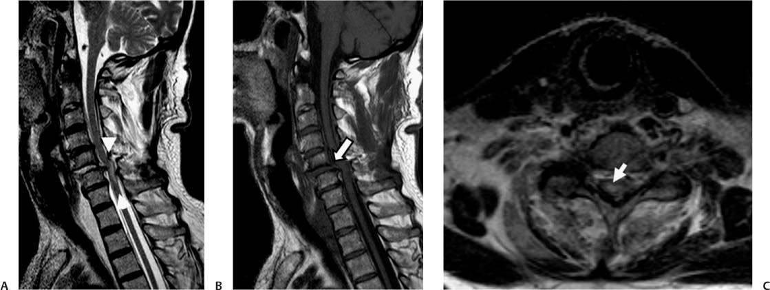
Case 49 A 37-year-old woman presenting with motor and sensory disturbances in the arms and legs after a motor vehicle accident. (A) Sagittal T2-weighted image (WI) shows subluxation and disk edema at C5-C6 with posterior osteophytes. Narrowing of the canal and edema of the cord (between arrowheads) are noted. (B) Sagittal T1WI shows subluxation and disk widening at C5-C6 (arrow). There are posterior osteophytes. (C) Note the central cord edema on the axial T2WI (arrow). No cord hemorrhage is demonstrated. • Cord contusion: Contusions tend to be severe in patients with degenerative changes. Magnetic resonance imaging (MRI) demonstrates increased cord signal and cord expansion. • Wallerian degeneration of the cord: This develops in the subacute phase (about 1 month) after spinal cord injury. MRI reveals hyperintense signal on T2WIs in the white matter tracts above (dorsal columns) and below (lateral columns) the level of the injury.
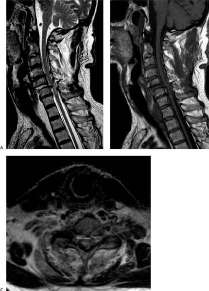
Clinical Presentation
Imaging Findings
Differential Diagnosis
Stay updated, free articles. Join our Telegram channel

Full access? Get Clinical Tree


