 Clinical Presentation
A human immunodeficiency virus–positive young adult man with progressive shortness of breath and cough.
 Imaging Findings
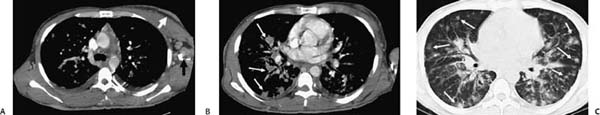
(A–C) Contrast-enhanced computed tomography of the chest: Mediastinal window images (A, B) demonstrate enlarged lymph nodes in the mediastinum and left axilla, as well as skin thickening and nodularity of the anterior chest wall (arrows, A). Abnormal peribronchovascular thickening and enlarged hilar lymph nodes, as well as hazy nodules scattered throughout the lung parenchyma, are better appreciated in the lung window image (arrows, C).
 Differential Diagnosis
• Kaposi sarcoma (KS): Bilateral pulmonary nodules in a peribronchovascular distribution associated with enhancing lymphadenopathy and skin thickening in a human immunodeficiency virus (HIV)–positive patient are suggestive of lung involvement from KS.
• Lymphoma:
Stay updated, free articles. Join our Telegram channel

Full access? Get Clinical Tree


