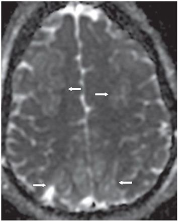
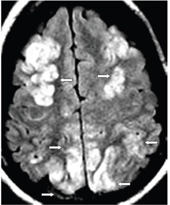
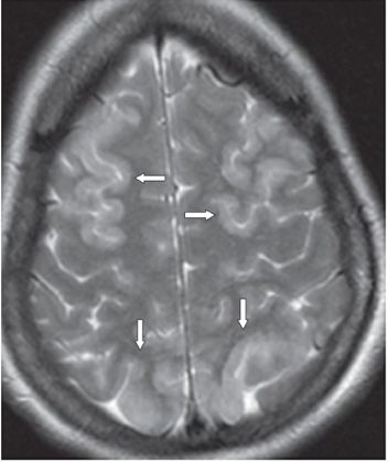
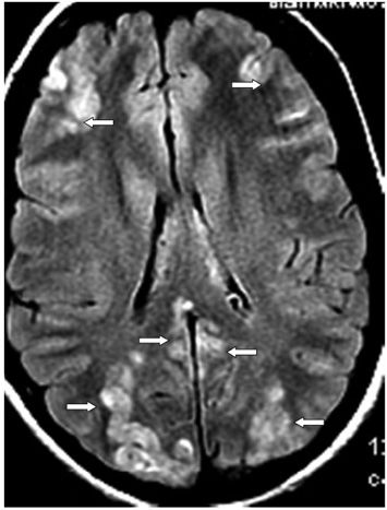
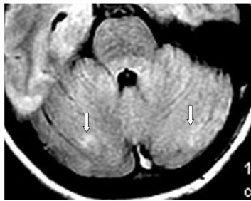
FINDINGS Figures 49-1. Axial DWI through the upper centrum semiovale showing patchy bilateral almost symmetrical frontoparietal cortical hyperintensity with corresponding ADC hyperintensity in Figure 49-2 (arrows). Figures 49-3 and 49-4. Axial FLAIR and T2WI, respectively, through same level confirm cortical patchy hyperintensity with mild effacement of sulci in the involved areas consistent with cortical swelling (arrows). Subcortical U fibers are intact. Figure 49-5. Axial FLAIR through the lateral ventricles. There is almost symmetrical cortical ribbon multifocal hyperintensity in bilateral frontal and parieto-occipital lobes (arrows). Temporal lobes (not shown) are also involved. Figure 49-6. Axial FLAIR through the cerebellum. There is patchy bilateral almost symmetrical cerebellar hyperintensity (arrows). Subtle changes are also present in the bilateral brachium pontes and tegmentum. Follow-up MRI 4 months after these images demonstrated complete resolution of the lesions.
DIFFERENTIAL DIAGNOSIS Posterior reversible encephalopathy syndrome (PRES), multifocal watershed subacute infarcts, multifocal cortical edema, acute demyelinating encephalomyelitis (ADEM), cerebral venous sinus thrombosis.
DIAGNOSIS Posterior reversible encephalopathy syndrome (PRES).
DISCUSSION
Stay updated, free articles. Join our Telegram channel

Full access? Get Clinical Tree


