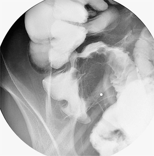
Case 49 A 35-year-old man presents to the gastroenterology clinic with right lower quadrant abdominal pain. Compression view from a small-bowel series shows nodularity (arrowheads) along the inferior aspect of the terminal ileum (TI), with sparing of the superior aspect. The cecum demonstrates mucosal irregularity along the medial aspect (arrows), with sparing of the lateral aspect. The result is a C-shaped configuration of the TI and cecum and a cone shape of the cecum. • Lymphoma: This is the top diagnosis, a classic cause of coned cecum with involvement of the TI. • Abscess from appendicitis or diverticulitis: This diagnostic option can produce mass effect and inflammatory changes at the TI and cecum. • Crohn disease and tuberculosis: These can also produce a coned cecum with TI involvement, but circumferential rather than eccentric involvement would be more common.
 Clinical Presentation
 Imaging Findings

 Differential Diagnosis
 Essential Facts
Stay updated, free articles. Join our Telegram channel

Full access? Get Clinical Tree


