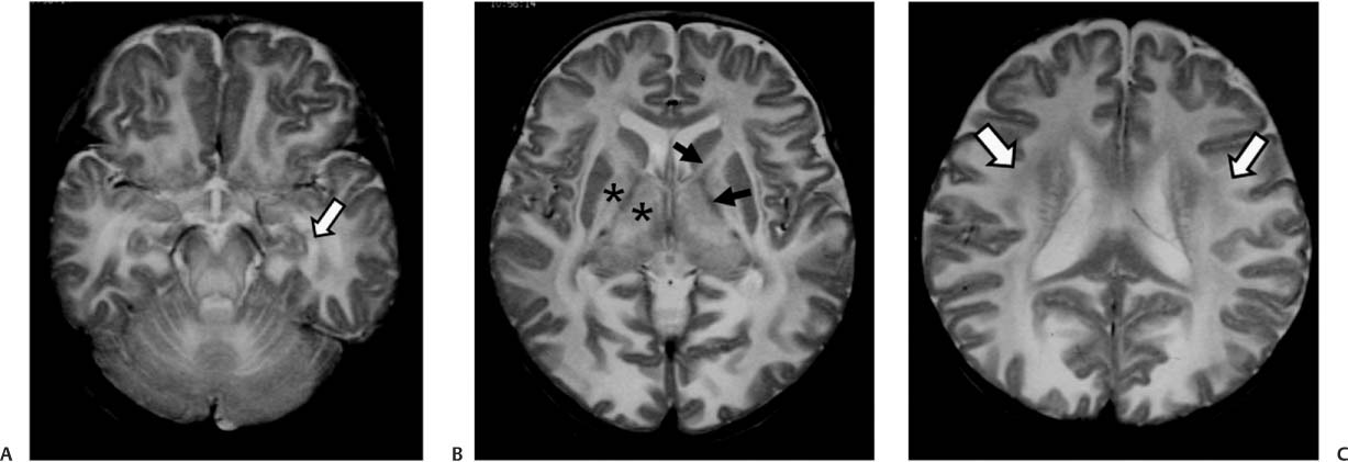
Case 5 An infant presenting with marked hypotonia, macrocephaly, and seizures. (A) Axial T2-weighted magnetic resonance (MR) image of the brain reveals an increased white matter T2 signal (arrow). (B) There is relative sparing of the internal capsule (arrows). Abnormal T2 signal is demonstrated in the thalami (asterisks) and globus pallidus. (C) Note the diffuse increase in the white matter T2 signal (arrows). • Canavan disease: This involves predominantly the sub-cortical U fibers. The occipital lobes are more involved than the frontal and parietal lobes. The thalami and basal ganglia are affected in severe cases.
Clinical Presentation
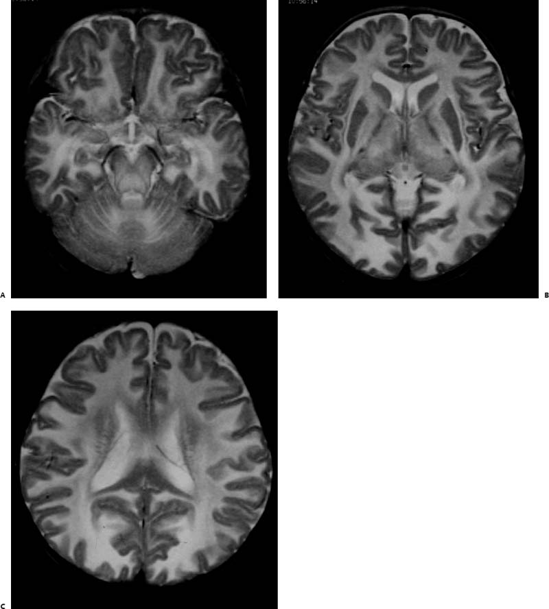
Imaging Findings
Differential Diagnosis
Stay updated, free articles. Join our Telegram channel

Full access? Get Clinical Tree


