 Clinical Presentation
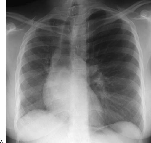
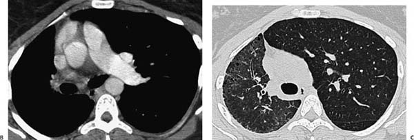
A 22-year-old woman with cough and dyspnea.
Further Work-up
 Imaging Findings
(A) Conventional chest radiograph shows asymmetric lung fields, with a smaller right hemithorax and ipsilateral deviation of the cardiac silhouette. (B, C) Contrast-enhanced computed tomography (CT) of the chest reveals absence of the right pulmonary artery and an asymmetric appearance of the lung parenchyma (white arrow, B). There are small, cystic-appearing changes in the periphery of the right lung, interlobular septal thickening, and a normal appearance of the left lung (black arrows, C).
 Differential Diagnosis
Stay updated, free articles. Join our Telegram channel

Full access? Get Clinical Tree


