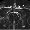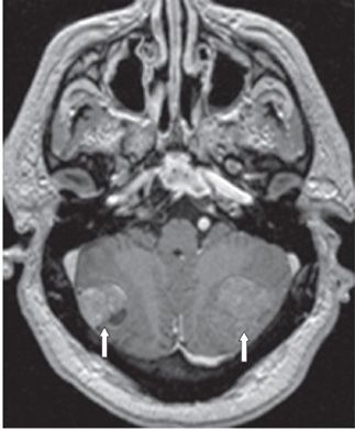
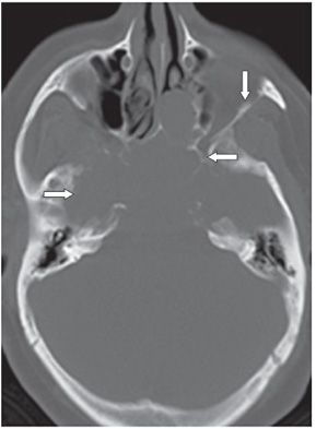
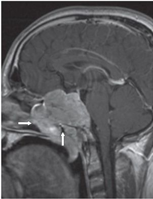
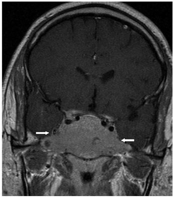
FINDINGS Figure 5-1. Sagittal post-contrast T1WI. There is a large inhomogeneous sellar mass with suprasellar extension and invasion of the sphenoid sinus (arrow). Figure 5-2. Two months later, a post-contrast axial T1WI shows bilateral cerebellar masses (arrows) that are metastases consistent with metastatic pituitary carcinoma. Figure 5-3. Axial NCCT in a companion case of invasive pituitary adenoma (PA) shows destruction of the central bony skull base (arrows). Figure 5-4. Sagittal post-contrast T1WI in the companion case. There is a large enhancing sellar mass with invasion of the sphenoid sinus and extension into the posterior nasal cavity (arrows). Figure 5-5. Coronal post-contrast T1WI shows invasion of both cavernous sinuses by the sellar mass (arrows).
DIFFERENTIAL DIAGNOSIS Meningioma, metastasis, clival chordoma, chondrosarcoma, plasmacytoma, malignant macroadenoma.
DIAGNOSIS Malignant macroadenoma.
DISCUSSION
Stay updated, free articles. Join our Telegram channel

Full access? Get Clinical Tree


