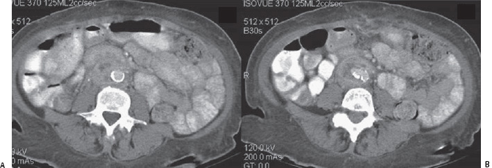
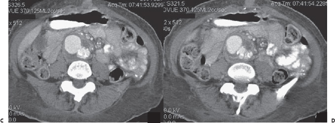
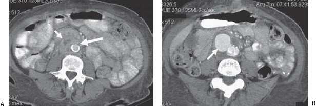
Case 5 The patient is a 68-year-old woman with chronic renal failure who is on dialysis. Further Work-up Image obtained 15 days later. (A) Axial image of the first contrast-enhanced computed tomographic (CT) scan shows a large area of soft-tissue density and irregular areas of decreased density surrounding the distal abdominal aorta and extending into the proximal portions of the iliac arteries (white arrowhead
 Clinical Presentation
 Imaging Findings
![]()
Stay updated, free articles. Join our Telegram channel

Full access? Get Clinical Tree


