Case 5
 Clinical Presentation
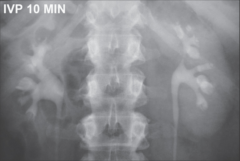
A 58-year-old man who has diabetes presents with acute bilateral flank pain and hematuria.
 Imaging Findings
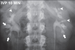
Ten-minute intravenous pyelogram (IVP) image of the kidney area shows that both kidneys are normal in size, shape, position, orientation, and outline. Many of the calices show extracaliceal collections of contrast, some of which are round (arrowheads), others flame-shaped (arrows). Opacified pelves and visualized upper ureters on both sides are normal in caliber and outline without filling defects.
 Differential Diagnosis
• Renal papillary necrosis: The characteristic features on IVP are contrast-filled cavities and fissures in the papillae. Various terms (e.g., “ball-on-T” “flame-shaped”) have been used to describe the various shapes of the extracaliceal contrast collections. In later stages, the papillae slough off. A sloughed papilla may cause a filling defect within the contrast-filled collecting system and ureter. The calices whose papillae have sloughed off have a flat or convex appearance.
• Renal tuberculosis: Multiple extracaliceal contrast collections are also found in renal tuberculosis. They are usually not limited to the renal papillae, are irregular, and may be associated with calcifications in the renal parenchyma.
• Hydronephrosis:
Stay updated, free articles. Join our Telegram channel

Full access? Get Clinical Tree


