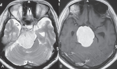
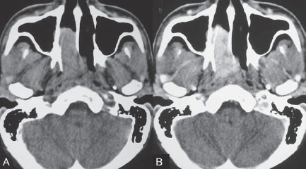
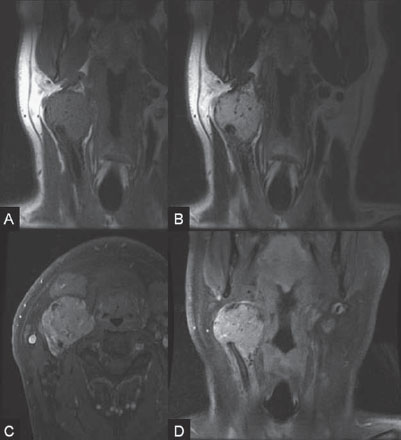
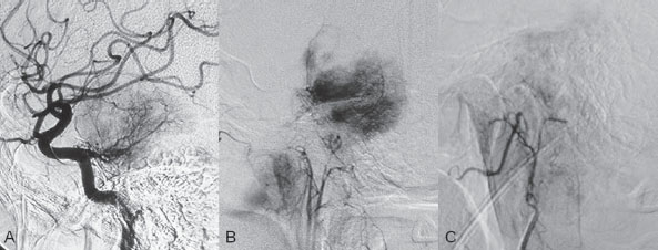
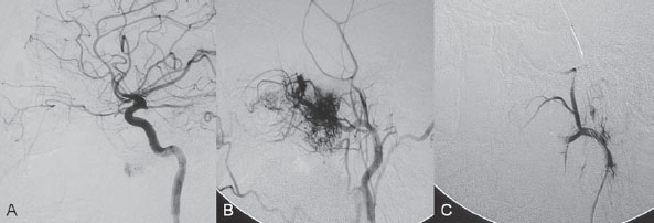
5 Tumors

PART VTumors


CASE 36


Case Description


Clinical Presentation


A 50-year-old woman has a history of headaches for nearly a year. One month before admission, she developed numbness of the right side of her face with ataxia. Physical examination shows decreased sensation in the territory of cranial nerve V, mild right-sided facial weakness, and ataxia. No motor weakness is present. MRI is performed, followed by angiography with preoperative embolization.


Image


Fig. 36.1 MRI. (A) T2-weighted and (B) post-gadolinium T1-weighted images.


Radiologic Studies


MRI


MRI revealed a well-defined extra-axial mass lesion at the right petroclival region with hypointense T1/hyperintense T2 signal and intense homogeneous enhancement on a post-contrast study. The lesion caused a significant mass effect on the right side of the pons and midbrain (Fig. 36.1). A linear, enhancing dural tail was observed along the clivus. The findings were consistent with the diagnosis of a petroclival meningioma.


DSA


The study demonstrated a hypervascular tumor blush, supplied predominantly by branches of the right meningohypophyseal trunk from the right internal carotid artery (ICA) and meningeal branches from the right neuromeningeal trunk of the right ascending pharyngeal artery (Fig. 36.2 A,B). Minimal supply from branches of the left meningohypophyseal trunk and left ascending pharyngeal artery was also observed.


Image


Fig. 36.2 DSA. (A) Right ICA and (B) right ascending pharyngeal artery angiograms in lateral view demonstrate a hypervascular tumor blush. (C) Angiogram of the right ascending pharyngeal artery after particle embolization shows obliteration of the supply to the tumor.


Diagnosis


Right petroclival meningioma


Treatment


EQUIPMENT


  • Standard 5F access (puncture needle, 5F vascular sheath)
  • Standard 5F multipurpose catheter (Guider Soft Tip; Boston Scientific, Natick, MA) with continuous flush and a 0.035-in hydrophilic guidewire (Terumo, Somerset, NJ)
  • A 0.018-in over-the-wire microcatheter (Prowler Select; Cordis, Warren, NJ) with a 0.014-in hydrophilic guidewire (Agility 14; Cordis)
  • Polyvinyl alcohol particles (Ivalon), 300 to 500 μm
  • Contrast material

DESCRIPTION


Following diagnostic angiography, a 5F multipurpose catheter was advanced into the proximal right external carotid artery (ECA). An over-the-wire microcatheter was introduced over a micro-guidewire into the neuromeningeal trunk of the right ascending pharyngeal artery. A mixture of 300- to 500- μm polyvinyl alcohol particles with contrast media was injected slowly under a blank roadmap until stagnation of the distal branches was observed. The microcatheter was removed, and the 5F multipurpose catheter was advanced into the proximal left ECA. The microcatheter was then placed into the left ascending pharyngeal artery, followed by particle embolization with the same technique. The microcatheter was removed, and the final control run showed obliteration of the external carotid supply to the tumor (Fig. 36.2C). Attempts to catheterize the meningohypophyseal trunks from the ICAs failed to reach a safe position for particle or glue embolization. The patient underwent surgical removal of the tumor within 24 hours after the procedure without any complications.


Discussion


Background


Meningiomas are extra-axial tumors arising from arachnoid cells of the meninges, which originate from the neural crest. They account for ~13 to 20% of all primary intracranial tumors and can be found at any location along the arachnoid membrane, most commonly along the cerebral convexity. The peak incidence is at 45 years of age, with a strong female predominance. Malignant lesions are very rare, but metastases may occur by seeding via the cerebrospinal fluid. Extraneural metastases to the lungs, liver, and lymph nodes may also occur. Because most meningiomas are benign and curable tumors, complete surgical resection remains the treatment of choice.


Noninvasive Imaging Workup


CT


  • More than 50% of meningiomas are hyperdense on unenhanced CT scans, and calcification can be seen in ~14%. Hypodensity surrounding the tumor is likely related to brain edema. Hyperostosis of the adjacent bone can also be seen on bone window.
  • Marked homogeneous enhancement with well-defined borders is typically seen on post-contrast CT.
  • Heterogeneous enhancement and tumor necrosis are unusual and suggest a more aggressive lesion.

MRI


  • Meningiomas are often homogeneously isointense to the brain parenchyma on T1-weighted sequences and hyperintense on T2-weighted sequences. Areas of flow void may be seen on T2weighted images, which may represent the highly vascular nature of these lesions and/or calcifications.
  • Contrast enhancement is necessary to determine the relationship of a meningioma to adjacent major cerebral arteries and veins.

CTA/MRA


  • The use of both CTA and dynamic MRA to determine the vascularity of meningiomas before pre-operative embolization has been reported.
  • Enlargement of the feeding arteries, typically from the middle meningeal artery for convexity lesions, can be demonstrated.

Invasive Imaging Workup


  • Angiography is usually not required for the diagnosis of a meningioma and is performed in most instances at the time of embolization.
  • The major arterial feeders to the center and dural base of a meningioma are typically from dural branches. In larger tumors, recruitment of pial supply from the anterior cerebral artery, middle cerebral artery, posterior cerebral artery, or cerebellar arteries to peripheral parts of the tumor may be seen.
  • Intraventricular meningiomas are supplied by the choroidal arteries.

Treatment Options


RADIOSURGERY


  • Radiosurgery has been increasingly used in recent years for meningiomas of the skull base. In small lesions, radiosurgery may be applied as a single modality or as an adjunctive treatment for residual tumor after surgical resection.
  • In our experience, the aim of radiosurgery is mainly to control tumor growth and recurrence. The size of the tumor usually does not significantly decrease after radiosurgery.

SURGICAL TREATMENT


  • Complete surgical resection is the best available treatment.
  • Various surgical methods have been used, depending on the location.

ENDOVASCULAR TREATMENT


  • Preoperative embolization can help reduce the vascularity of the tumor and therefore decrease blood loss and facilitate complete removal of the tumor during surgery.
  • As in the presurgical management of arteriovenous malformations, embolization can be performed to occlude those feeding arteries that will be difficult to reach at surgery. It can also be used to devascularize the tumor bed and induce shrinkage and necrosis, thereby facilitating removal at surgery. The superselectivity required and the embolic material chosen will be different in these two circumstances.
  • Delay of surgery for up to 3 weeks after embolization to allow time for maximum tumor necrosis, which softens the tumor and facilitates complete removal, has been reported.
  • Although some studies have preferred the use of smaller particles (i.e., 45–150 μm) for maximum tumor necrosis, in our experience, particles of 300 to 500 μm are safer to avoid penetration through the skull base and intratumoral anastomoses with subsequent emboli and ischemic stroke. However, if larger particles are used, as in our case, the optimal surgical timing is within 24 hours.
  • The use of permanent liquid embolic agents (i.e., glue and Onyx) has been reported for the preoperative embolization of meningiomas; in our opinion, both should be used with caution because of their ability to penetrate the above-mentioned anastomoses.
  • The dural branches arising from the ECAs, usually the middle meningeal artery, are embolized first. The tip of the microcatheter should ideally be placed within or adjacent to the tumor bed. If a good, far-distal position within the arterial feeders from the dural branches of the ICA or vertebral artery (VA) can be obtained, these will subsequently be embolized. In cases of aggressive lesions, recurrent tumors, or young patients, the pial supply may also be embolized to improve the chance for complete resection.
  • Swelling of a tumor after embolization may occur very rarely, usually within 48 to 72 hours of the procedure, and may be treated with steroids. This type of complication does not appear to occur when the tumor bed is satisfactorily embolized but may reflect incomplete embolization when there is dual supply from both pial and dural vasculature.

Possible Complications


  • Hemorrhage may be intratumoral or may be epidural, subdural, or subarachnoidal when it is due to arterial perforation.
  • Stroke or cranial nerve palsies may result from distal embolization through the skull base and intratumoral anastomotic channels.
  • Swelling of the tumor may occur after embolization.

Published Literature on Treatment Options


Many studies in previous literature have shown benefits of the preoperative embolization of meningiomas, especially larger ones, to help reduce blood loss during surgery, time required for surgical resection, and length of hospital stay. Blood loss is significantly reduced if the tumor is devascularized more than 90%. In the past, it was generally accepted that the optimal timing for surgery after embolization to reduce the blood loss was within 24 hours; however, this concept has changed in the past years. Several authors have used permanent liquid embolic agents (glue and Onyx), which makes it possible to delay surgery until maximum tumor necrosis and softening occur. This phenomenon also occurs with the use of smaller particles, which penetrate and occlude the capillary bed of the tumor; when these embolic agents are used, the optimal interval between embolization and surgery for tumor softening is 7 to 9 days. The complication rates for the preoperative embolization of meningiomas vary among institutions but are generally accepted to be between 5 and 7%. A significant risk factor for complications is the use of smaller particles or liquid embolic agents. The use of a temporary balloon has been advocated to reduce the chance of stroke during the embolization of branches of the ICAs and VAs.


The balloon can be positioned across the origin of the feeding dural vessel after it has been superselectively catheterized. The balloon is inflated while the embolization is performed via the microcatheter with either particles or liquids. Another method is to position the balloon distal to the tumor-feeding dural branch and inflate the balloon while embolization is performed in the main parent vessel ((ICA, VA) through a microcatheter positioned immediately adjacent to the origin of the tumor-feeding dural branch. Flow into the dural branch must be significant for the embolic material to reach this vessel while the balloon is inflated for distal protection. Once flow to the tumor has greatly diminished, the parent vessel is “cleaned” with repeated injections of saline and either removal of the mixture of blood and saline or intentional flushing of the column down to the external carotid system. The balloon system is then deflated and removed. Obviously, anticoagulation is necessary under these circumstances


PEARLS AND PITFALLS__________________________________________________


  • Most meningiomas are benign tumors, and therefore complete surgical resection is the best treatment modality.
  • The major arterial supply to the central tumor and dural attachment is through the meningeal arteries, which in most cases originate from the ECA and can be embolized with a low risk for complications.
  • Smaller particles are associated with a higher risk for complications, and liquid embolic agents (e.g., glue and Onyx) should be used carefully because of their ability to penetrate through the skull base and intratumoral anastomoses.
  • As in the embolization of arteriovenous malformations, the tip of the microcatheter should ideally be placed as close as possible to the tumor bed. At that point, liquid material or small particles can be gently injected for deeper penetration of the tumor.

Further Reading


Dowd CF, Halbach VV, Higashida RT. Meningiomas: the role of preoperative angiography and embolization. Neurosurg Focus 2003;15(1):E10


Engelhard HH. Progress in the diagnosis and treatment of patients with meningiomas. Part I: diagnostic imaging, preoperative embolization. Surg Neurol 2001;55(2):89–101


Lasjaunias P, Berenstein A, terBrugge K. Surgical Neuroangiography. Vol 2: Clinical and Endovascular Treatment Aspects in Adults. 2nd ed. New York, NY: Springer-Verlag; 2004


Tymianski M, Willinsky RA, Tator CH, Mikulis D, TerBrugge KG, Markson L. Embolization with temporary balloon occlusion of the internal carotid artery and in vivo proton spectroscopy improves radical removal of petrous-tentorial meningioma. Neurosurgery 1994;35(5):974–977, discussion


CASE 37


Case Description


Clinical Presentation


An 18-year-old man presents with a history of recurrent episodes of epistaxis for 3 months and progressive stuffiness of the right nostril. His neurologic examination is normal. CT is performed, followed by angiography with preoperative embolization.


Image


Fig. 37.1 (A) Unenhanced and (B) post-contrast axial CT scans at the level of the nasopharynx.


Radiologic Studies


CT


CT showed a slightly hyperdense mass at the right posterior nasal cavity involving the nasal septum and extending into the nasopharynx. Marked, slightly heterogeneous enhancement was seen on a post-contrast study (Fig. 37.1). Given the age and sex of the patient, these findings led to the diagnosis of a juvenile angiofibroma.


DSA


The study revealed a hypervascular tumor blush, supplied predominantly by branches of the right internal maxillary artery and ascending palatine artery from the right facial artery (Fig. 37.2 A,B). Minimal supply from the mandibular artery of the right internal carotid artery (ICA) through the foramen lacerum was also observed.


Diagnosis


Right-sided juvenile angiofibroma


Image


Fig. 37.2 DSA. (A) Right ICA and (B) right internal maxillary artery angiograms in lateral view. (C) Post-particle embolization angiogram of the right internal maxillary artery shows obliteration of the supply to the tumor.


Treatment


EQUIPMENT


  • Standard 5F access (puncture needle, 5F vascular sheath)
  • Standard 5F multipurpose catheter (Guider Soft Tip; Boston Scientific, Natick, MA) with continuous flush and a 0.035-in hydrophilic guidewire (Terumo, Somerset, NJ)
  • A 0.021-in over-the-wire microcatheter (Prowler Select Plus; Cordis, Warren, NJ) with a 0.014-in hydrophilic guidewire (Agility 14; Cordis)
  • Polyvinyl alcohol particles (Ivalon), 355 to 500 μm
  • Contrast material

DESCRIPTION


Following diagnostic angiography, a 5F multipurpose catheter was advanced into the proximal right external carotid artery. An over-the-wire microcatheter was introduced over a micro-guidewire into the distal internal maxillary artery beyond the origin of the middle meningeal artery. A mixture of 355- to 500-μm polyvinyl alcohol particles with contrast media was injected slowly under a blank roadmap until stagnation of the distal branches was observed. The microcatheter was then advanced into the right ascending palatine artery of the right facial artery, followed by particle embolization with the same technique. The microcatheter was removed, and the final control run showed obliteration of the external carotid supply to the tumor (Fig. 37.2C). The patient underwent further surgical removal of the tumor within 24 hours after the procedure without any complications.


Discussion


Background


Juvenile angiofibromas are relatively uncommon tumors of vascular origin that are histologically benign but locally invasive. They account for ~0.5% of all head and neck tumors, typically developing in male adolescents. The peak age is ~14 to 17 years. The clinical symptoms of a juvenile angiofibroma are related directly to its size and extension. The most common are nasal obstruction and epistaxis. The tumor typically originates in the posterior nasal cavity and usually extends to the pterygopalatine fossa. Occasionally, it may arise from the nasal septum. The classification system of Fisch is often employed for staging juvenile angiofibromas; stages I and II tumors are still localized to the nasal cavity, nasopharynx, and pterygopalatine fossa, whereas stages III and IV tumors involve the infratemporal fossa, orbit, and skull base. Complete surgical resection is the best treatment choice; however, local recurrence may be seen in up to 35% of cases. Malignant transformation is extremely rare and in most cases reported in the literature is secondary to radiation therapy.


Noninvasive Imaging Workup


CT


  • An intense, rather homogeneously enhancing soft-tissue mass can be identified on unenhanced and post-contrast CT scans. Extension through the pterygopalatine fossa with subsequent expansion is a common finding.
  • Bone windows are useful to detect small areas of bone erosion by the tumor.

MRI


  • Juvenile angiofibromas usually have an intermediate signal on T1-weighted sequences, compared with the high signal of fat and low signal of muscle, and they have a high signal on T2-weighted sequences. Small flow voids may also be seen on T2-weighted sequences, which are related to the vascularity of the tumor.
  • Intense contrast enhancement can be visualized on post-gadolinium T1-weighted sequences, and coronal and sagittal views are necessary for the evaluation of intracranial extension and skull base involvement.

CTA/MRA


  • CTA and MRA are not routinely used for the evaluation of juvenile angiofibromas.
  • Enlargement of the internal maxillary arteries, which are the major feeding arteries, may be seen. In stages III and IV with skull base involvement, narrowing of the ICAs due to encasement by the tumor may be visualized.

Invasive Imaging Workup


  • Angiography is not required for the diagnosis of a juvenile angiofibroma and is performed only before embolization.
  • The extracranial portion of the tumor is supplied mainly by branches of the internal maxillary artery, the accessory meningeal artery, the ascending pharyngeal artery, and intrapetrous and intracavernous branches of the ICA, which extend extracranially through the neural foramina.
  • Tumors with orbital and skull base extension may recruit additional supply from the ethmoidal branches of the ophthalmic artery and intracranial branches of the intracavernous ICA.

Treatment Options


RADIOSURGERY


  • Although the use of radiation alone for the treatment of advanced juvenile angiofibromas (stages III and IV) has been reported, we still believe that radiosurgery should be reserved for cases with residual tumor after surgery because of the possibility of secondary malignant transformation, secondary tumor formation, and other late complications related to radiation.

SURGICAL TREATMENT


  • Complete surgical resection is the best available treatment.
  • In general, for stages I and II tumors, endoscopic surgery is usually sufficient, and preoperative embolization may not be necessary. A multimodality approach is usually employed for stages III and IV tumors, in which preoperative embolization is followed by radical surgery and possible radiosurgery for residual intracavernous tumor.

ENDOVASCULAR TREATMENT


  • Preoperative embolization can help reduce the vascularity of the tumor and therefore decrease blood loss and facilitate complete removal of the tumor during surgery.
  • The supplying vessels from the external carotid artery, including the internal maxillary artery, accessory meningeal artery, ascending palatine artery from the facial artery, and ascending pharyngeal arteries, are the major targets of preoperative embolization.
  • Particles are the preferred embolic material, and the particle size should be larger than 150 to 200 μm to avoid penetration through the skull base and intratumoral anastomoses. Studies have also shown that because of the presence of micro-arteriovenous shunts within the tumoral bed, the use of smaller particles may result in the formation of pulmonary emboli. In our experience, we usually use 355- to 500-μm polyvinyl alcohol particles for tumor devascularization, followed by Gelfoam strip or tube particles to close the proximal feeding artery.
  • The optimal time for surgery is within 24 hours after embolization.
  • Percutaneous puncture and direct embolization with liquid agents such as glue or Onyx have been reported for juvenile angiofibromas that are supplied mainly by branches of the ICA; however, experience with this technique is necessary because it carries a significantly higher risk for embolic complications to the brain caused by retrograde penetration of the embolic material through tumoral anastomoses into the ICA.
  • In cases with extensive involvement of the skull base, sacrifice of the ICA after a balloon occlusion test may facilitate complete removal of the tumor.

Possible Complications


  • Stroke, cranial nerve palsies, or blindness may result from distal embolization through the skull base and intratumoral anastomotic channels.

Published Literature on Treatment Options


Preoperative embolization is currently generally accepted as the standard management of juvenile angiofibromas, particularly large tumors with skull base extension (Fisch stages III and IV). In smaller tumors (stages I and II), several studies have shown that preoperative embolization helps to decrease blood loss during endoscopic surgery, the duration of packing, and length of hospital stay. Although several reports have been published about the direct percutaneous puncture and intratumoral embolization of juvenile angiofibromas, this technique requires significant operator experience to avoid the potentially devastating complications of ICA stroke.


PEARLS AND PITFALLS__________________________________________________


  • Juvenile angiofibromas are benign, locally aggressive vascular tumors that occur predominantly in male adolescents.
  • Complete surgical resection is the best treatment modality and can be facilitated by preoperative embolization, especially in patients with large lesions.
  • Smaller particles are associated with a higher risk for complications because of their ability to penetrate through the skull base and intratumoral anastomoses, causing stroke or cranial nerve palsy, and through intratumoral micro-arteriovenous shunts, causing pulmonary emboli.

Further Reading


Andrade NA, Pinto JA, Nobrega MdeO, Aguiar JE, Aguiar TF, Vinhaes ES. Exclusively endoscopic surgery for juvenile nasopharyngeal angiofibroma. Otolaryngol Head Neck Surg 2007;137(3):492–496


Casasco A, Houdart E, Biondi A, et al. Major complications of percutaneous embolization of skull-base tumors. AJNR Am J Neuroradiol 1999;20(1):179–181


Valavanis A, Christoforidis G. Applications of interventional neuroradiology in the head and neck. Semin Roentgenol 2000;35(1):72–83


CASE 38


Case Description


Clinical Presentation


A 42-year-old man feels a slowly growing mass in his neck below the angle of the mandible. On examination, a nontender mass is palpated in the carotid space that is mobile in the lateral plane but immobile in the craniocaudal plane. There are no signs of pain or dysphagia, and the patient does not experience syncope. Contrast-enhanced MRI is performed.


Image


Fig. 38.1 MRI of the neck. (A) T1 pre-contrast, (B) T1 post-contrast, (C) axial fat-suppressed T2, and (D) coronal T1 post-contrast fat-suppressed sequences demonstrate an intensely enhancing T2-hyperintense lesion with intralesional flow voids at the carotid bifurcation. This lesion separates the ECA and ICA.


Radiologic Studies


MRI


MRI demonstrated a densely enhancing T2-hyperintense lesion in direct proximity to the right neurovascular bundle with intratumoral flow voids and separation of the external and internal carotid arteries (Fig. 38.1). No other tumors were visible.

Only gold members can continue reading. Log In or Register to continue

Stay updated, free articles. Join our Telegram channel

Dec 23, 2015 | Posted by in INTERVENTIONAL RADIOLOGY | Comments Off on 5 Tumors

Full access? Get Clinical Tree

Get Clinical Tree app for offline access