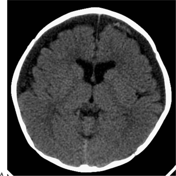
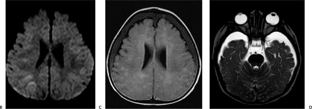
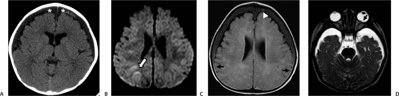
Case 50 A 7-month-old child with a history of vomiting. Retinal hemorrhages were found at the clinical examination. (A) Computed tomography (CT) of the brain demonstrates subdural collections in both frontal convexities, with mixed attenuation (asterisks). (B) Diffusion-weighted magnetic resonance imaging (MRI) shows restricted diffusion in the right parietal subcortical white matter (arrow). (C) Axial fluid-attenuated inversion recovery image shows hematocrit effect, with blood elements layering in the dependant portion of the collection (arrows) and focal areas of high signal within the subdural space anteriorly, consistent with acute blood (arrowhead). (D) Note the presence of retinal hemorrhages in the posterior pole of the left globe (arrow).
Clinical Presentation
Further Work-up
Imaging Findings
Differential Diagnosis
Stay updated, free articles. Join our Telegram channel

Full access? Get Clinical Tree


