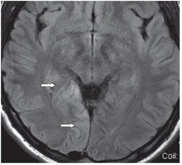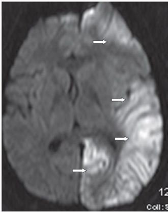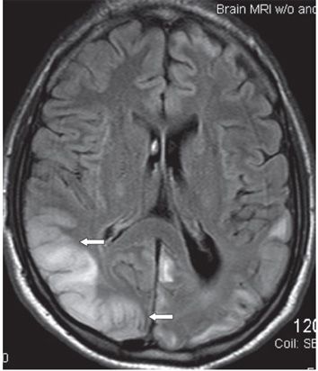


FINDINGS Figure 50-1. Axial MRI FLAIR through the occipital lobes. There is a left medial occipital lobe smudgy hyperintensity mainly cortical/subcortical in location (arrows) at the time of initial presentation with seizures. Two months later, he suffered another set of seizures. Figure 50-2. An axial FLAIR image from that time showing a right medial occipital lobe cortical/subcortical smudgy hyperintensity and sulcal effacement (arrows). There is resolution of initial left occipital hyperintensity. Figure 50-3. Axial DWI through the level of the thalamus 2 years after Figure 50-2 following new seizures. There is circumferential left hemispheric cortical/subcortical area of restricted diffusion (arrows) and smudgy hyperintensity with sulcal effacement and mass effect on FLAIR (not shown). There is subtle mild midline shift from left to right. Nine months later he returned with new episodes of seizures. Figure 50-4. Axial FLAIR through the occipital lobes demonstrates a new right parietal, temporal, and occipital cortical/subcortical smudgy T2 hyperintensity with sulcal effacement (arrows). There is restricted diffusion in these regions. There is residual hyperintensity and volume loss (dilated left lateral ventricle and sulci) in the left hemisphere.
DIFFERENTIAL DIAGNOSIS
Stay updated, free articles. Join our Telegram channel

Full access? Get Clinical Tree


