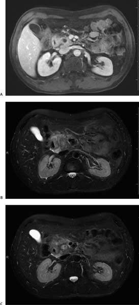
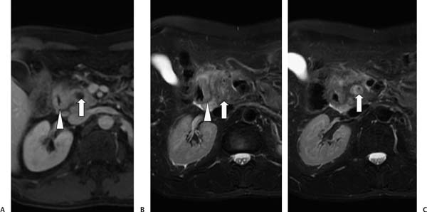
Case 50 A 40-year-old man presents with recurrent epigastric pain and a 20-lb weight loss. (A) T1-weighted magnetic resonance image (MRI) shows a focal lesion of low signal intensity (arrow) in the pancreatic head. There is focal thickening of the adjacent wall of the second portion of the duodenum (arrowhead). (B) T2-weighted image shows heterogeneous signal intensity in the pancreatic head (arrow) and focal duodenal wall thickening (arrowhead). There is increased signal in the peripancreatic tissues and in the pancreaticoduodenal groove, consistent with fluid and edema. (C) The focal lesion in the pancreatic head (arrow) is heterogeneously bright on T2. • Focal pancreatitis:
 Clinical Presentation
 Imaging Findings
 Differential Diagnosis
![]()
Stay updated, free articles. Join our Telegram channel

Full access? Get Clinical Tree


