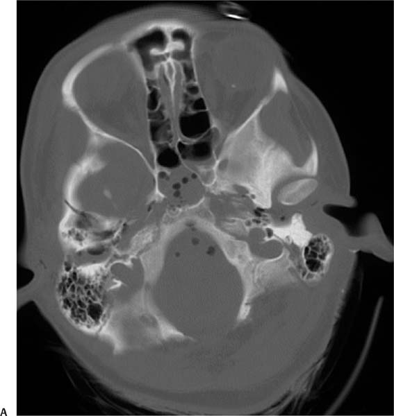
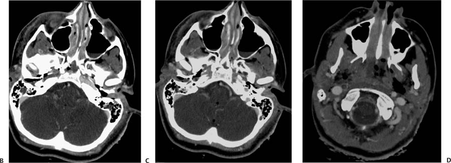
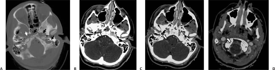
Case 51 A 36-year-old woman injured in a motor vehicle collision. (A) Computed tomography (CT) of the brain with bone windows shows a complex fracture of the skull base involving the sphenoid and temporal bones (black arrows) and compromise of the anterolateral wall of the right carotid canal (white arrow). (B) Axial image from a CT angiogram (CTA) of the brain shows absent opacification of the petrous right internal carotid artery (ICA) secondary to traumatic vascular injury (arrow). (C) Axial image from a CTA of the brain shows absent opacification of the petrous right ICA secondary to traumatic vascular injury (arrow). Pneumocephalus and neck emphysema are also noted. (D) Axial CTA at the level of C1 shows absent flow in the right ICA secondary to traumatic vascular injury (arrow
Clinical Presentation
Further Work-up
Imaging Findings
![]()
Stay updated, free articles. Join our Telegram channel

Full access? Get Clinical Tree


