 Clinical Presentation
A 35-year-old woman who is a smoker with the gradual onset of mild dyspnea.
Further Work-up
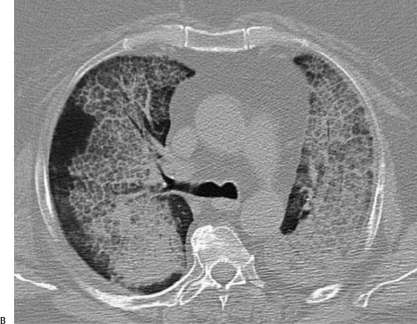
 Imaging Findings
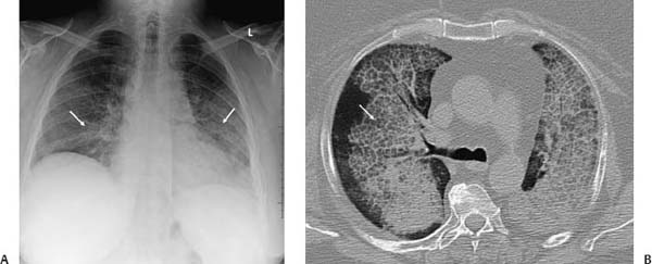
(A) Chest radiograph demonstrates central ground-glass and linear interstitial opacity bilaterally (arrows). (B) Contrast-enhanced computed tomography (lung windows) shows geographic ground-glass opacity (arrow), with interlobular septal thickening sharply demarcated from normal lung.
Stay updated, free articles. Join our Telegram channel

Full access? Get Clinical Tree


