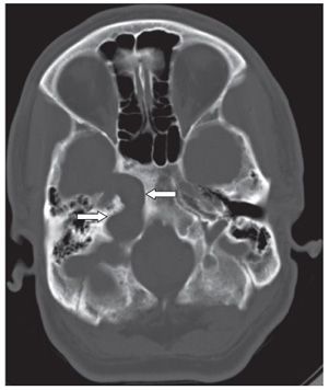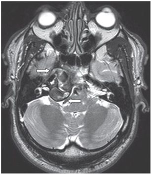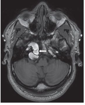


FINDINGS Figure 51-1. Axial NCCT through the petrous apices, brain window. There is a well-marginated isodense mass (with streak artifacts) centered on the right petrous apex bulging into the middle cranial fossa and posterior fossa right cerebellopontine angle (CPA) (arrows) with mass effect on surrounding structures. Figure 51-2. Axial NCCT at the level of foramen magnum. There is chronic smooth bony remodeling of the right petrous apex and adjoining clivus (arrows). Figure 51-3. Axial T2WI through the mass. There is a heterogeneous intensity pattern within the well-defined mass. There is no surrounding edema. Figure 51-4. Axial T1WI through the mass. The mass is homogeneously hyperintense (arrow).
DIFFERENTIAL DIAGNOSIS
Stay updated, free articles. Join our Telegram channel

Full access? Get Clinical Tree


