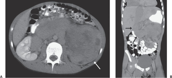
Case 51 A 3-year-old child with a palpable left-sided abdominal mass. (A,B) Axial and coronal reformat images of a computed tomography (CT) abdominal examination demonstrate a large, heterogeneous mass in the left renal fossa, with abnormal dilatation and a continuous filling defect in the left renal vein, inferior vena cava (IVC), and inferior part of the right atrium (black and white arrows). In addition, there is a large soft-tissue mass in the base of the right hemithorax (arrowhead). • Wilms tumor:
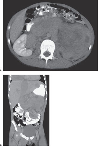
Clinical Presentation
Imaging Findings
Differential Diagnosis
![]()
Stay updated, free articles. Join our Telegram channel

Full access? Get Clinical Tree


