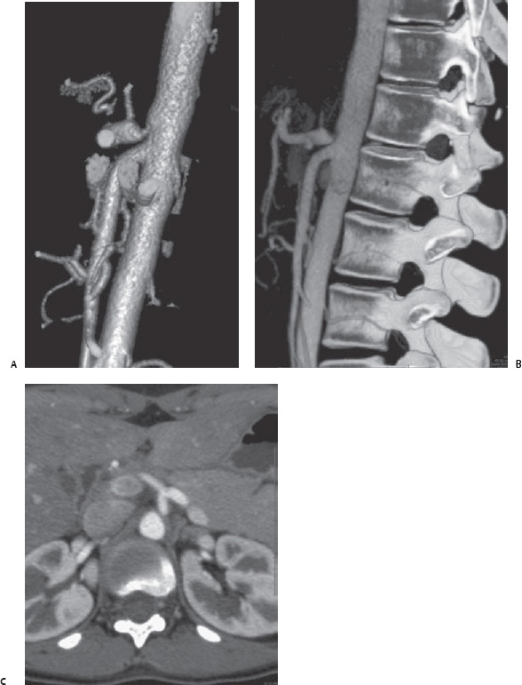
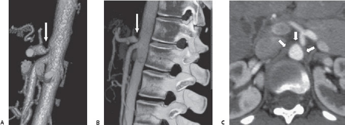
Case 51 A 16-year-old patient presents with chronic abdominal pain and weight loss of 20 lbs. (A) A three-dimensional volume-rendered computed tomographic angiogram (CTA) obtained during exhalation accentuates partial extrinsic compression (arrow) of the superior aspect of the celiac artery. Annotated is the left gastric artery (arrowhead). (B) Sagittal reformatted image obtained during exhalation again shows the extrinsic compression (arrow) of the superior aspect of the celiac artery. (C) Sagittal reformatted image obtained during inhalation shows resolution of the extrinsic compression (arrow).
 Clinical Presentation
 Essential Facts
 Differential Diagnosis
Stay updated, free articles. Join our Telegram channel

Full access? Get Clinical Tree


