CASE 51
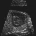
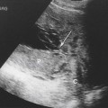
Used with permission from McGahan JP, et al: Fetal abdomen and pelvis. In McGahan JP, Goldberg BB [eds]: Diagnostic Ultrasound, 2nd ed. New York: Informa Healthcare USA, 2008; 1278.
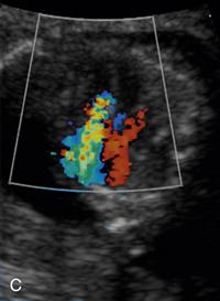
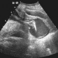
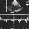
Used with permission from McGahan JP, et al: Fetal abdomen and pelvis. In McGahan JP, Goldberg BB [eds]: Diagnostic Ultrasound, 2nd ed. New York: Informa Healthcare USA, 2008; 1278.
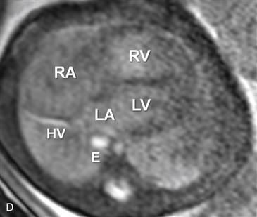
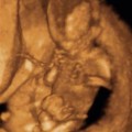
RV = right ventricle; RA = right atrium, LA = left atrium; LV = left ventricle; HV = hepatic vein; E = esophagus. Used with permission from Anderson Publishing Ltd. From Hellinger J, et al. Fetal MRI in the Third Dimension. Applied Radiology. 2010; 39(7)8-19. © Anderson Publishing Ltd.
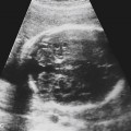
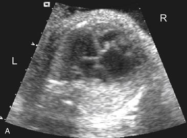
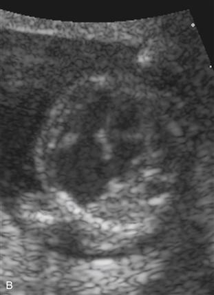
History: A pregnant woman in the late second trimester presents for a routine ultrasound study.