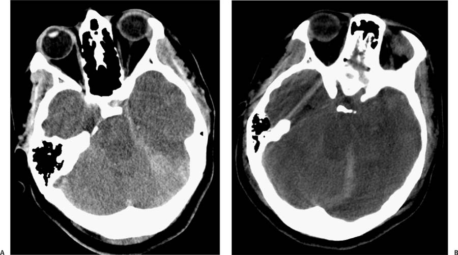
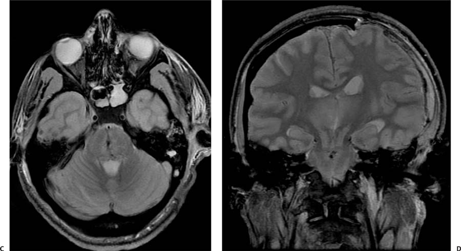
Case 52 A 26-year-old who was involved in a car accident and is now comatose. (A) Axial computed tomography (CT) of the brain shows signs of cerebral edema, with low attenuation of the brain and loss of distinction between gray and white matter. effacement of the basal cisterns (arrows) and deformity of the midbrain are evident. (B) Axial CT demonstrates effacement of the perimesencephalic cistern (arrows) and deformity of the midbrain, with reduced transverse diameter and elongation in the anteroposterior direction. Note a subdural hematoma over the left tentorium. (C) Axial gradient-echo T2*-weighted image (WI) reveals petechial hemorrhages in the central pons (arrow). (D) Coronal T2WI shows downward displacement of the thalami and midbrain (asterisk). The pontine hemorrhages are again demonstrated.
Clinical Presentation
Further Work-up
Imaging Findings
Stay updated, free articles. Join our Telegram channel

Full access? Get Clinical Tree


