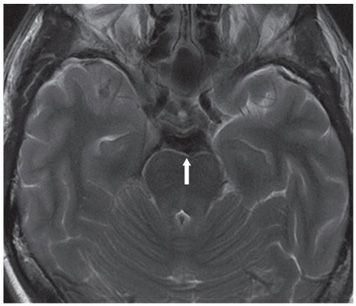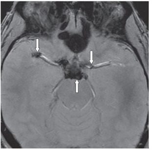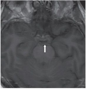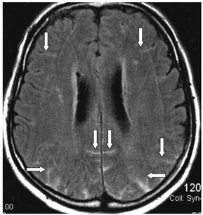


FINDINGS Figure 52-1. Axial NCCT through the posterior fossa. There is subarachnoid space (SAS) hyperdensity in the posterior and middle cranial fossae (arrow). Figure 52-2. Axial T2WI through the suprasellar cistern. There is diffuse hypointensity in the SAS (arrow). Figure 52-3. Axial GRE through the suprasellar cistern. There is diffuse hypointensity/signal void in the suprasellar cistern and lateral fissures (arrows). Figure 52-4. Axial non-contrast T1WI through the suprasellar cistern. There is diffuse isointensity in the suprasellar cistern (arrow). Figure 52-5. Axial FLAIR through the lateral ventricles in a companion patient with SAH. There is bilateral scattered sulcal hyperintensity (arrows).
DIFFERENTIAL DIAGNOSIS N/A.
DIAGNOSIS Acute subarachnoid hemorrhage (ASAH).
DISCUSSION
Stay updated, free articles. Join our Telegram channel

Full access? Get Clinical Tree


