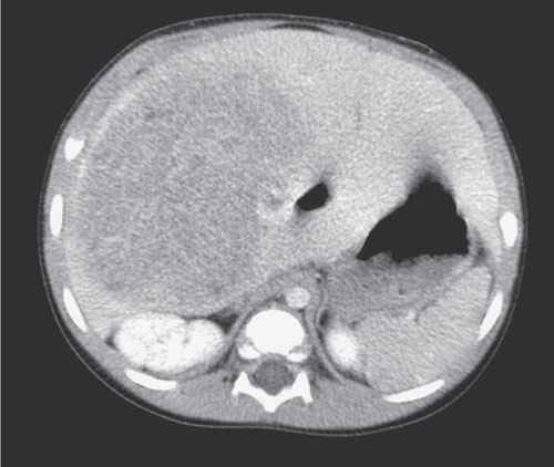
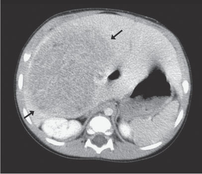
Case 52 A 1-year-old with a right-sided abdominal mass. Axial computed tomography image obtained after the intravenous administration of contrast demonstrates a large, heterogeneous, hypoattenuating mass occupying most of the liver (arrows). • Hepatoblastoma:
Clinical Presentation
Imaging Findings
Differential Diagnosis
![]()
Stay updated, free articles. Join our Telegram channel

Full access? Get Clinical Tree


