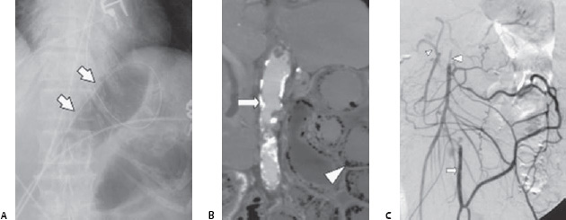
Case 52 A 78-year-old woman with a history of postprandial pain presents with intense abdominal pain. (A) Radiograph shows dilated loops of bowel and possible pneumatosis (arrows). (B) Contrast-enhanced computed tomography (CT) shows pneumatosis (arrowhead) and aor tic calcifications (arrow). (C) Inferior mesenteric artery (IMA: arrow) angiogram shows superior mesenteric artery (SMA: large arrowhead) stenosis and reconstitution via the marginal artery from the IMA; the celiac artery is presumed stenotic because its branches are reconstituted via pancreaticoduodenal arteries supplied by the SMA (proper hepatic artery: small arrowhead).
 Clinical Presentation
 Imaging Findings
 Differential Diagnosis
Stay updated, free articles. Join our Telegram channel

Full access? Get Clinical Tree


