 Clinical Presentation
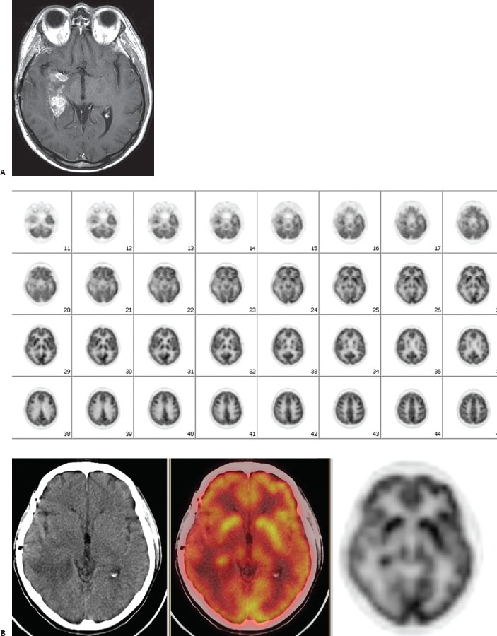
A 48-year-old man with glioblastoma multiforme status post resection and radiation therapy presents for restaging.

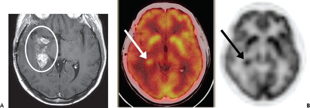
(A) Axial T1 postcontrast brain MRI demonstrates abnormal enhancement in the medial right temporal lobe (circle). (B) Axial brain FDG-PET and a selected hybrid PET/CT image demonstrate increased radiotracer accumulation in the right temporal lobe, most prominently in the posterior medial aspect (arrows). This corresponds with the enhancing region on MRI. The CT portion shows low density in this area, indicating edema.
 Differential Diagnosis
• Tumor recurrence: An enhancing lesion with increased FDG accumulation indicates recurrent tumor, given the history.
• Radiation necrosis:


Stay updated, free articles. Join our Telegram channel

Full access? Get Clinical Tree


