 Clinical Presentation
A 62-year-old man with shortness of breath and a history of asbestos exposure.
 Imaging Findings
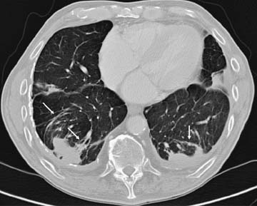
Contrast-enhanced computed tomography (lung window) through the lung bases shows bilateral pleural thickening and calcified pleural plaques. Foci of consolidation abut the posterior pleura bilaterally. Long curvilinear opacities extend from the masses anteriorly (arrows). The margins of the masses form an acute angle with the pleura.
 Differential Diagnosis
Stay updated, free articles. Join our Telegram channel

Full access? Get Clinical Tree


