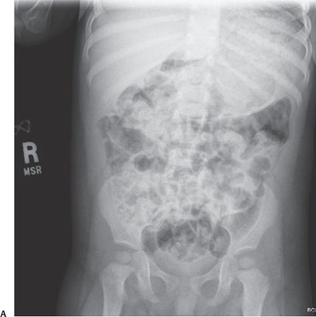
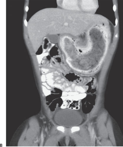
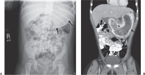
Case 53 A young girl with abdominal pain and an epigastric mass. (A) Abdominal radiograph demonstrates a heterogeneous soft-tissue mass in the shape of the stomach (arrows). (B) Coronal reformat computed tomography image of the abdomen after oral and intravenous administration of contrast demonstrates a large, heterogeneous, hypodense gastric filling defect (arrow) that conforms to the shape of the stomach and is surrounded by oral contrast material, indicating that it does not arise from the gastric wall. It contains hypodense pockets of gas.
Clinical Presentation
Further Work-up
Imaging Findings
Stay updated, free articles. Join our Telegram channel

Full access? Get Clinical Tree


