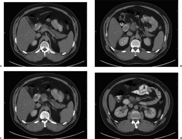
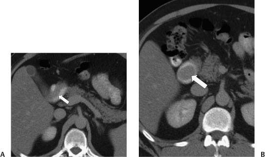
Case 53 A 47-year-old man presents to the gastroenterology clinic with abdominal pain and recurrent gastrointestinal bleeding. (A) Contrast-enhanced computed tomography (CT) shows a soft-tissue filling defect (arrow) originating from the immediate postbulbar duodenal wall. No adjacent wall thickening and no lymphadenopathy are present. (B) The soft-tissue filling defect (arrow) is polypoid and terminates in the distal descending duodenum. • Adenoma: This is the most likely diagnosis as it is commonly solitary and periampullary in origin. • Brunner gland hamartoma: This is also solitary and well circumscribed, in the proximal duodenum, and either homogeneous or heterogeneous on CT, depending on the amount of cystic degeneration and fat. • Lymphoma: This is a third option. It may be polypoid, but this presentation is less common than invasive lymphoma.
 Clinical Presentation
 Imaging Findings
 Differential Diagnosis
Stay updated, free articles. Join our Telegram channel

Full access? Get Clinical Tree


