 Clinical Presentation
A 70-year-old woman who is a smoker with chronic cough.
Further Work-up
 Imaging Findings
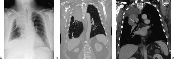
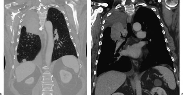
(A) Chest radiograph demonstrates right upper lobe atelectasis (arrow). The minor fissure is elevated and medially displaced. Proximally, however, there is convexity of the fissure. (B) Coronal computed tomography (CT) reconstruction (lung windows) demonstrates the right upper lobe atelectasis. A pulmonary nodule is seen at the right base that was not convincingly evident on the radiograph (arrow, B). (C) Coronal CT reconstruction (soft-tissue windows) clearly depicts a proximal mass obstructing the right upper lobe bronchus (arrow, C).
 Differential Diagnosis
Stay updated, free articles. Join our Telegram channel

Full access? Get Clinical Tree


