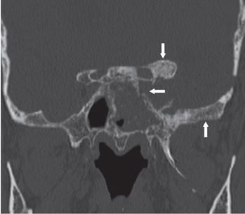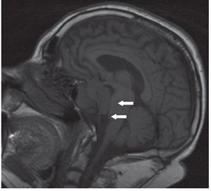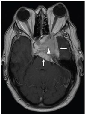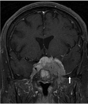


FINDINGS Figure 54-1. Axial NCCT through the sella turcica. There is a mild-to-moderate hyperdense mass occupying the left parasellar region with linear calcifications in its lateral margin (arrow). Figure 54-2. Coronal bone window NCCT through the anterior clinoid processes. There is hyperostosis especially of the left anterior clinoid process and greater wing of the sphenoid bone (vertical arrows) and lytic areas in the left sphenoid sinus wall (transverse arrow). There is a left sphenoid sinus opacification and osteoneogenesis. Figure 54-3. Sagittal non-contrast T1WI shows a large isointense homogeneous sella mass that erodes the clivus and extends posteriorly into the interpeduncular and pre/pontine cisterns (arrows). Figure 54-4. Axial post-contrast T1WI through the mass. There is an avidly enhancing mass centered in the left parasellar region and extends to the sella turcica. The mass compresses the left temporal lobe laterally (transverse arrow) and the brainstem posteriorly (vertical arrow). The left internal carotid artery (ICA) is narrowed and encased by the mass (triangle). There is a left orbital proptosis. Figure 54-5. Coronal post-contrast T1WI through the sella. There is invasion of both cavernous sinuses and the sella by the mass. There is a left middle cranial fossa floor dural tail (arrow).
DIFFERENTIAL DIAGNOSIS
Stay updated, free articles. Join our Telegram channel

Full access? Get Clinical Tree


