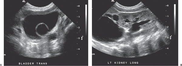
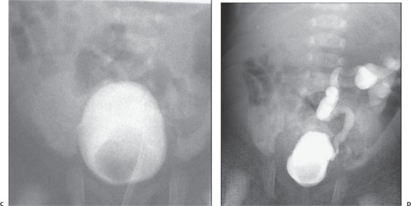
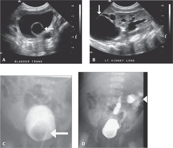
Case 54 An infant with a recent history of urinary tract infection. (A,B) A transverse sonogram through the urinary bladder demonstrates a cystic structure adjacent to the expected location of the left ureterovesical junction (arrow), almost certainly representing a ureterocele, and a longitudinal sonogram of the left kidney demonstrates mild lower pole hydronephrosis and severe upper pole hydronephrosis (arrow). (C,D) Two radiographic images from a voiding cysto-urethrogram demonstrate a filling defect in the bladder, corresponding to the ureterocele seen on sonography (arrow), as well as a grade 4 reflect into the lower pole of the left kidney (arrowhead), which is inferiorly displaced by the hydronephrotic (obstructed) upper pole.
Clinical Presentation
Further Work-up
Imaging Findings
Stay updated, free articles. Join our Telegram channel

Full access? Get Clinical Tree


