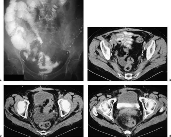
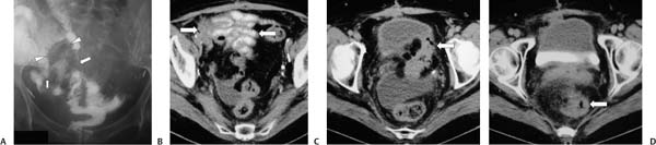
Case 54 A 62-year-old woman presents with abdominal pain. (A) Barium study shows fixed and separated small bowel (arrowheads), acute angulation of small bowel (small arrow), thickened folds, and stricture formation (large arrow). (B) Computed tomography (CT) shows multiple fixed, matted enteric loops (arrows) with adjacent soft-tissue infiltration, suggesting fibrosis. Bowel wall and fold thickening is also evident. (C,D) More caudal images show obliteration of the fat planes normally separating pelvic structures and rectosigmoid involvement (arrows). • Chronic radiation enteritis: This is the top diagnosis, given the abdominal clips from prior surgery, tethering and angulation of small bowel, and involvement of nonadjacent structures, suggesting a distribution within a radiation portal (rectosigmoid colon, bladder, and small bowel). • Crohn disease:
 Clinical Presentation
 Imaging Findings
 Differential Diagnosis
![]()
Stay updated, free articles. Join our Telegram channel

Full access? Get Clinical Tree


