Case 54
Case History
A 65-year-old woman has right breast microcalcifications that have been stereotactically mammographically biopsied and are malignant. She now presents for needle localization of these microcalcifications before lumpectomy.
Physical Examination
• normal exam
Mammogram
Mass (Fig. 54–1)
• margin: indistinct
• shape: irregular
• density: equal density
Calcifications
• type: fine linear/branching
• distribution: grouped/clustered
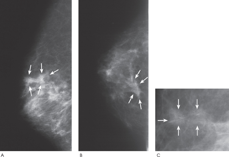
Figure 54–1. In the upper inner quadrant, there is a cluster of fine linear calcifications. These calcifications are associated with a dumbbell-shaped ill-defined mass (arrows). The shape of this mass and the subtle multinodular pattern of the surrounding parenchyma suggest that the tumor may be multifocal. (A). Right ML mammogram. (B). Right CC mammogram. (C). Right MLO magnification mammogram.
Ultrasound
Frequency
• 10 MHz
Mass
• margin: ill defined
• echogenicity: hypoechoic


Stay updated, free articles. Join our Telegram channel

Full access? Get Clinical Tree


