 Clinical Presentation
A 53-year-old man with fever and shortness of breath 2 weeks after stem cell transplant.
Further Work-up
 Imaging Findings
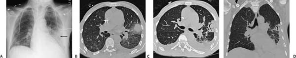
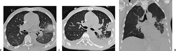
(A) Frontal chest radiograph demonstrates air-space opacity in the periphery of the left lung (arrow). (B) Noncontrast computed tomography (CT; lung window) demonstrates a solid mass in the left upper lobe with surrounding ground-glass opacity (arrow). There is patchy ground-glass opacity in the superior segments of the lower lobes. A left pleural effusion is present. (C) Noncontrast CT (lung window) obtained 1 week later demonstrates a peripheral crescentic collection of air surrounding the central solid mass (black arrow). The surrounding ground-glass opacity has resolved. (D) Noncontrast CT (coronal) shows the air crescent (black arrow) to advantage.
 Differential Diagnosis
• Invasive aspergillosis:
Stay updated, free articles. Join our Telegram channel

Full access? Get Clinical Tree


