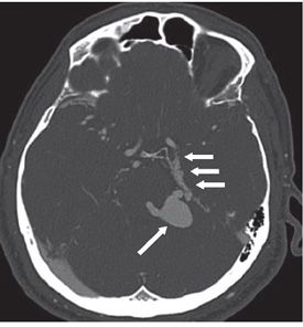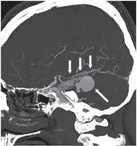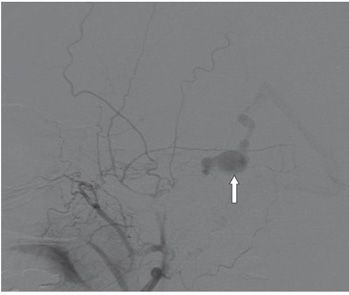


FINDINGS Figure 55-1. NCCT through the dorsum sella. There is asymmetrically increased density along the left petroclinoid ligament and anterior tentorium edge (transverse arrows). There is a more focal appearing hyperdensity slightly lateral and posterior to the left ambient cistern (vertical arrow) associated with a medial tubular density overlying the midbrain. Figures 55-2 and 55-3. CT angiography with axial and sagittal MIP reconstructions. There are numerous small caliber vascular structures extending from the left petroclinoid ligament posteriorly along the left medial edge of the tentorium (short white arrows). There is a large venous varix (long white arrow) adjacent to the left midbrain and pons corresponding to the hyperdensity seen on the NCCT. Figure 55-4. Digital subtraction angiography (DSA). Left internal maxillary artery injection. The DAVF is supplied by the middle meningeal artery with early shunting into the varix (arrow) and drainage into the vein of Galen and straight sinus. Tiny meningeal feeders are seen corresponding to the CTA findings in Figure 55-3.
DIFFERENTIAL DIAGNOSIS Dural arteriovenous fistula (DAVF)/malformation. Pial arteriovenous malformation (AVM), aneurysm.
DIAGNOSIS DAVF/malformation.
DISCUSSION
Stay updated, free articles. Join our Telegram channel

Full access? Get Clinical Tree


