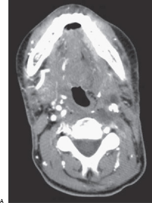
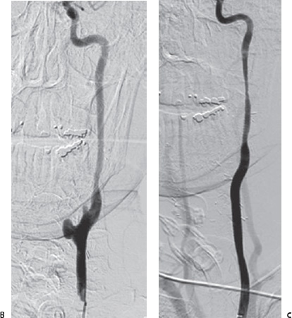
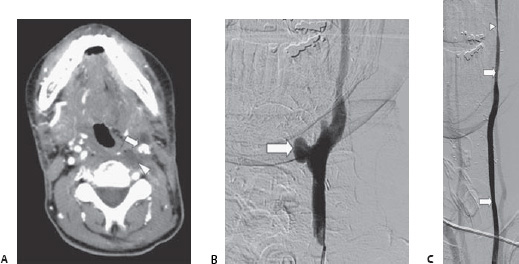
Case 55 A 47-year-old man who has head and neck cancer previously treated with radiation to the neck presents to the emergency department with bleeding into the mouth. Further Work-up (A) Contrast-enhanced computed tomographic (CT) scan of the neck 3 days before carotid rupture shows an irregularly marginated left carotid artery (arrow) passing through necrotic neck tumor (arrowhead). (B) Conventional angiogram in the frontal projection shows pseudoaneurysm (arrow
 Clinical Presentation
 Imaging Findings
![]()
Stay updated, free articles. Join our Telegram channel

Full access? Get Clinical Tree


