Case 55
Case History
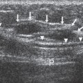
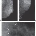
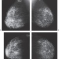
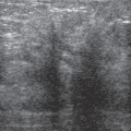
A 57-year-old woman with an new right breast dimpling. Mammographically, two suspicious abnormalities are identified: lesion A in the medial inferior breast (which corresponds to the skin dimpling) and mass B at 12:00. Unfortunately, only the 12:00 mass (B) is adequately localized for biopsy. The medial inferior abnormality (A) is not confidently identified during an attempt to perform mammographic stereotactic biopsy and is not localized on the initial breast sonogram. Lesion A is only identified sonographically after breast MRI demonstrates the mass.
Physical Examination
• right breast: subtle skin dimpling in medial breast associated with vague firmness
• left breast: normal exam
Mammogram
Mass (Fig. 55–1)
• margin: indistinct
• shape: irregular
• density: equal density
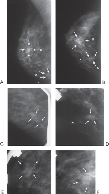
Figure 55–1. The skin dimpling in the medial inferior quadrant (marked by a radiopaque dot) is associated with an ill-defined mass (A), which is better identified on the CC spot compression (D). Lesion A appears as a subtle density associated with architectural distortion in Figure 55–1
Stay updated, free articles. Join our Telegram channel

Full access? Get Clinical Tree


