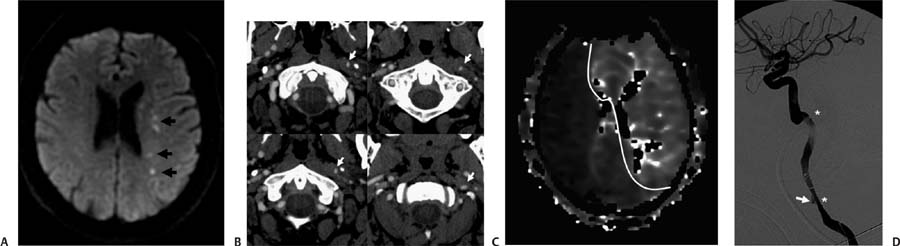
Case 56 A 55-year-old man with the sudden onset of right-sided weakness and neck pain. (A) Axial Diffusion-weighted image (WI) demonstrates multiple punctate foci of restriction in the left centrum semiovale, consistent with acute watershed infarcts (arrows). (B) Serial axial images from a computed tomography (CT) angiogram show narrowing of the left internal carotid artery (ICA) with a rotating flat lumen (arrows). (C) CT perfusion time-to-peak image demonstrates delayed cerebral blood flow in the territory of both anterior cerebral arteries and the left middle cerebral artery (delineated). (D) Digital subtraction angiography shows dissection of the left ICA from its origin, with a long segment of narrowing (between asterisks) and a spiral configuration of the lumen. There is a pseudoaneurysm in the proximal aspect of the dissection (arrow). • Carotid dissection:
Clinical Presentation
Further Work-up
Imaging Findings
Differential Diagnosis
![]()
Stay updated, free articles. Join our Telegram channel

Full access? Get Clinical Tree


