 Clinical Presentation
A 43-year-old man with chest pain.
Further Work-up
 Imaging Findings
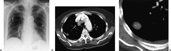
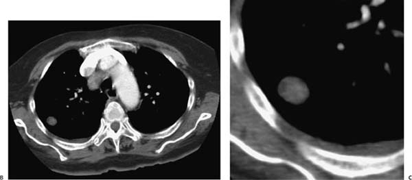
(A) Chest radiograph demonstrates a well-circumscribed 2-cm pulmonary nodule in the right upper lobe (arrow). (B) Contrast-enhanced computed tomography (CT) confirms a well-circumscribed solitary pulmonary nodule of heterogeneous density (arrow). (C) Magnified CT image of the nodule demonstrates a focus of macroscopic fat in its lateral aspect (arrow).
 Differential Diagnosis
• Hamartoma: Fat density within a solitary pulmonary nodule is a reliable indicator of a pulmonary hamartoma.
• Granuloma:
Stay updated, free articles. Join our Telegram channel

Full access? Get Clinical Tree


