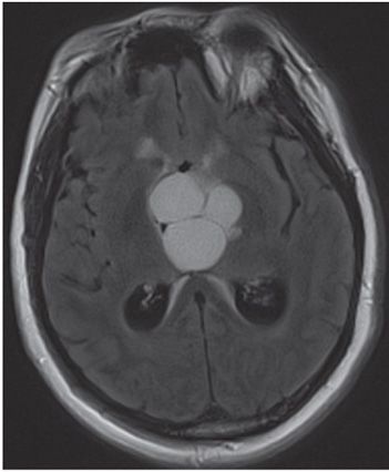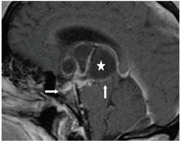

FINDINGS Figure 56-1. Axial NCCT through the suprasellar region. There is a suprasellar predominantly cystic mass with central calcifications (transverse arrow). There is dilation of the trigones (vertical arrow) consistent with hydrocephalus. Figure 56-2. Axial FLAIR through the suprasellar region. There is a hyperintense multicystic lobulated mass in the suprasellar region probably due to blood/protein in the fluid. Figure 56-3. Post-contrast sagittal T1WI. The mass is multiloculated (star) with enhancement of the cyst walls and septations with compression of the third ventricle and the midbrain (vertical arrow). The sella turcica is normal (transverse arrow), indicating suprasellar location of the mass.
DIFFERENTIAL DIAGNOSIS Rathke cleft cyst, Craniopharyngioma (CP), suprasellar arachnoid cyst, hypothalamic astrocytoma, pituitary adenoma, dermoid or epidermoid tumors, thrombosed aneurysm, and germ cell tumors.
DIAGNOSIS Craniopharyngioma (CP).
DISCUSSION
Stay updated, free articles. Join our Telegram channel

Full access? Get Clinical Tree


