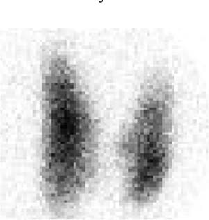
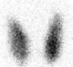
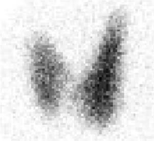
CASE 56 A 34-year-old woman presents with a possible right thyroid nodule palpated by the referring physician. Her thyroid function test values are normal. Fig. 56.1 Right anterior oblique pinhole collimator view, 123I. Fig. 56.2 Anterior pinhole collimator view, 123I. Fig. 56.3 Left anterior oblique pinhole collimator view, 123I. • 0.300 mCi of 123I administered orally 4 hours before uptake and scan • Five-minute pinhole collimator images in right anterior oblique, anterior, and left anterior oblique projections • An appropriately shielded sodium iodide thyroid probe is positioned with the crystal surface 25 cm from the surface being measured. Obtain counts of the following for 2 minutes: • Calculate the iodine uptake with the following formula: The 24-hour uptake of radioiodine is 20%. Three views (Figs. 56.1, 56.2, and 56.3) demonstrate homogeneous tracer distribution throughout the thyroid gland. The pyramid-shaped gland has smooth contours and relatively less activity at the periphery, reflecting thinner tissue there. A faint isthmus unites the right and left lobes. Each normal thyroid lobe measures approximately 5 cm in length and 2 cm in width. On physical examination, the gland feels normal in size (estimated weight, 15–30 g), configuration, and consistency. No nodule is palpated. • Normal thyroid gland Normal thyroid gland. Diagnosis final, no clinical follow-up. Thyroid scintigraphy is clinically important for the diagnosis and treatment of a variety of benign and malignant thyroid disorders, including the evaluation of goiters, differential diagnosis of hyperthyroidism, and function of thyroid nodules. Optimal thyroid scintigraphy requires an understanding of the two common imaging agents, radioactive 123I and 99m
Clinical Presentation
Technique
 Thyroid bed
 Thigh (body background)
 Room background
 Pill standard (in a neck phantom)

Image Interpretation
Differential Diagnosis
Diagnosis and Clinical Follow-Up
Discussion
![]()
Stay updated, free articles. Join our Telegram channel

Full access? Get Clinical Tree


