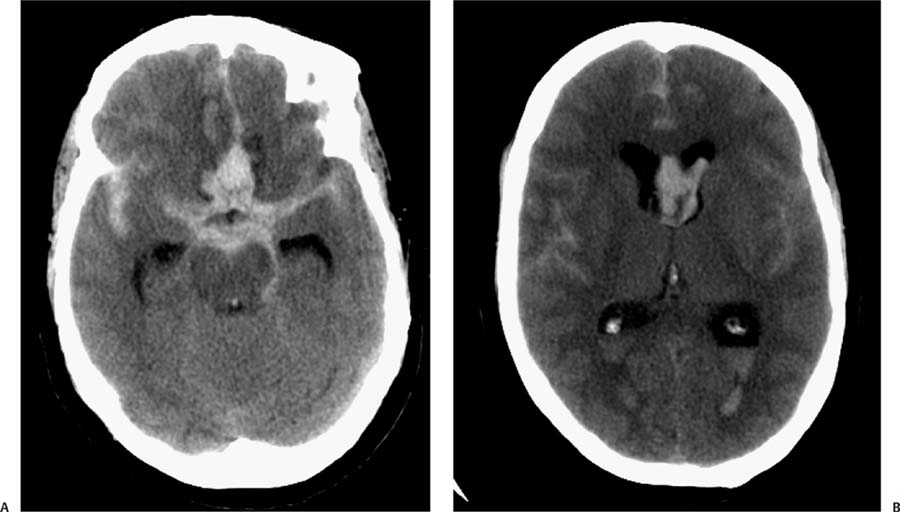
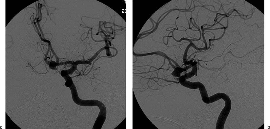
Case 57 A young woman who has fallen from her own height after “the worst headache of her life.” (A) Axial nonenhanced computed tomography (CT) shows subarachnoid hemorrhage (SAH) in the basal cisterns that is more prominent in the interhemispheric region. Areas of low attenuation in the gyri recti are noted, consistent with edema or ischemia (arrows). There is also intraventricular hemorrhage. (B) Axial nonenhanced CT shows SAH in the interhemispheric and sylvian regions. There is also intraventricular hemorrhage (arrows). (C) Oblique projection of a digital subtraction angiogram (DSA) of the left internal carotid artery (ICA) reveals a lobulated aneurysm in the anterior communicating artery (arrow). There is no narrowing of the vessels to suggest vasospasm. (D) Lateral projection of a DSA of the left ICA shows a lobulated aneurysm in the anterior communicating artery (arrow). • Ruptured cerebral aneurysm:
Clinical Presentation
Further Work-up
Imaging Findings
Differential Diagnosis
![]()
Stay updated, free articles. Join our Telegram channel

Full access? Get Clinical Tree


