 Clinical Presentation
A 44-year-old woman with uveitis.
Further Work-up
 Imaging Findings
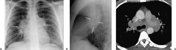
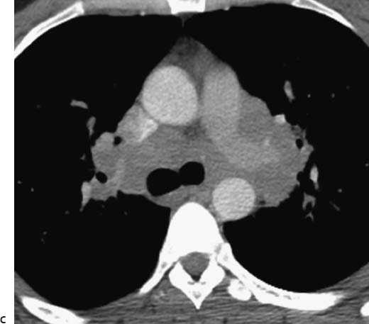
(A) Posteroanterior chest radiograph demonstrates symmetric hilar and right paratracheal lymphadenopathy (arrows). The lungs are normal. (B) Lateral chest radiograph shows the lymphadenopathy to advantage (arrows). (C) Computed tomography (CT) of the chest (soft-tissue windows) confirms substantial mediastinal and hilar lymphadenopathy (arrows).
 Differential Diagnosis
• Sarcoidosis: Symmetric hilar and mediastinal lymphadenopathy in a patient with uveitis is suggestive of sarcoidosis.
• Lymphoma: Lymphadenopathy due to lymphoma is typically asymmetric and abuts the cardiac margins. Uveitis is not expected.
• Tuberculosis (TB): In primary TB, the lymphadenopathy is asymmetric and ipsilateral to the pulmonary consolidation. This patient’s symptoms are not consistent with TB.
Stay updated, free articles. Join our Telegram channel

Full access? Get Clinical Tree


